Cloning Techniques in Antibody Discovery and Engineering
Cloning techniques are an essential tool in antibody discovery and engineering. These techniques involve the insertion of DNA fragments encoding antibodies or antibody fragments into a vector or plasmid, which can then be used to express the proteins of interest in a host organism. The ability to clone and change DNA fragments has changed the field of antibody engineering. This has made it possible to make new antibodies with better properties and more uses in therapy. Similarly, other technologies such as hybridomas, PCR, peptide and protein displays, and Next Generation sequencing have all contributed to the development and optimization of recombinant monoclonal antibodies1.
Cloning is used to produce a large collection of antibodies, improve the affinity and specificity of antibodies, and engineer antibodies for use in medicine. By using cloning techniques, researchers can produce a large number of antibody variants and screen them for desirable properties. Additionally, cloning techniques can be used to modify existing antibodies or create novel antibody formats with improved pharmacokinetic and pharmacodynamic properties.
Cloning often involves the use of plasmids, which are tiny DNA molecules packaged in circular form and may be found in bacteria. They are readily replicable in bacteria and may be altered to carry genes of interest, such as genes for antibodies. In addition, they can be changed to have genes.
Types of cloning techniques
Several types of cloning techniques are commonly used in antibody discovery and engineering, including restriction cloning, PCR cloning, Gateway cloning, Gibson Assembly, Golden Gate cloning, In-fusion cloning and TOPO Cloning.
Restriction Cloning
Cloning by restriction is one of the most commonplace and easy strategies used for antibody discovery and engineering. The technique involves the use of restriction enzymes to cut both the plasmid and the DNA fragment to be cloned. The DNA fragment is then inserted into the plasmid using DNA ligase.
Restriction cloning is used in the antibody discovery process to clone antibody genes into expression vectors so that the antibodies themselves may be expressed and produced. Cloning with genetic material restriction is a method that is both economical and easily accessible. On the other hand, it is unsuitable for cloning big DNA fragments and performs poorly in high-throughput applications.
Commercial vectors, protocols, and bacterial research systems are available for this approach. Restriction enzymes with various overhangs and recognition sites are also commonly available2.

PCR Cloning
PCR cloning is a widely used technique in molecular biology that enables researchers to amplify specific DNA fragments or genes for further analysis or manipulation. PCR, a polymerase chain reaction, amplifies DNA sequences by utilizing polymerase enzymes3. The polymerase chain reaction (PCR) is the foundation of this method. PCR is a fast and accurate way to generate a large number of identical copies of a target DNA sequence.
Once the gene or DNA fragment of interest is amplified using PCR, it can be inserted into a plasmid vector using a variety of cloning techniques such as restriction enzyme digestion or ligation. These vectors are typically modified to include antibiotic resistance markers or other selectable markers that allow for easy selection and isolation of the cloned DNA.
PCR cloning is often preferred over other cloning methods for applications that require high throughput, as it is a rapid and efficient way to generate large quantities of DNA fragments or genes. Due to its practicality, quickness, and need for only a tiny quantity of starting material, this approach is perfect for cloning uncommon or low-expressed antibodies.
Additionally, PCR cloning can be used to create site-directed mutagenesis or to add tags or other sequences to a gene of interest, allowing researchers to study gene function or protein expression in greater detail. There have been a number of other methods created as alternatives to PCR cloning, such as recombinase-dependent cloning, ligation independent cloning (LIC), TA cloning, and PCR-mediated cloning4.
Gateway Cloning
Gateway cloning is a method that allows DNA segments to be cloned into target vectors without the use of restriction enzymes as a prerequisite5. It is a highly efficient cloning technique and involves the use of site-specific recombination. The antibody gene or DNA fragment is first cloned into an entry vector, which contains attL sites. The entry vector is then recombined with a destination vector containing attR sites, which results in the insertion of the antibody gene into the destination vector.
Despite gateway cloning being a quick and effective cloning technique, the cost of recombination enzymes and gateway vectors might be high. Following gateway cloning, it is also rather difficult to transition to some other relevant cloning method, like restriction cloning6.
Gibson Assembly
Using a mix of enzymes and DNA polymerase, the Gibson assembly method enables the assembling several DNA fragments into a single piece of DNA7. Thus, overlapping DNA fragments can be used to assemble, for example, a complete antibody gene. The DNA fragments are annealed using a mixture of DNA polymerase, exonuclease, and ligase. This technique is particularly useful for the generation of complex antibody libraries.
Gibson cloning makes it easy to assemble numerous DNA segments in the desired orientation without undesired sequences at the junction. Any double-stranded dsDNA fragment, such as a PCR product or manufactured oligo, may be easily ligated plasmid backbone if correctly constructed8.
Golden Gate cloning
Golden Gate cloning is a method of restriction cloning that involves cutting DNA at predetermined locations using enzymes belonging to the Type IIS restriction family9.
Golden gate cloning has two benefits: Firstly, since the resultant overhangs will be unique and maintain the orientation of the cloning process, the whole cloning phase (both digestion and ligation) may be completed in a single reaction using a single Type IIS restriction enzyme. Secondly, the restriction site is transcribed on the insert and vector in a way that ensures no unwanted sequence being left in the finished product as well as elimination of all recognition motifs. The method has been applied by scientists in order to produce suitably tailored overhangs, which can then be joined together in an effective manner10.
In-fusion cloning
In-fusion cloning allows the insertion of DNA fragments into linearized plasmids by using a mix of enzymes and DNA polymerase11. It involves the use of PCR to generate overlapping DNA fragments that are assembled using a recombinase-based method. In-Fusion Cloning is a very effective and ligation-independent method of cloning that is based on the fusing of complementary ends of an insert and vector.
This method makes it possible to clone any desired gene into any vector/carrier at any locus in a simple manner using only a single step. This technique is particularly useful for the generation of antibody libraries12.
For large-scale research projects that require the expression of thousands of genes as proteins, an effective technique for cloning genes into protein expression vectors is crucial. However, conventional cloning techniques that rely on restriction enzyme digestion and ligation are not practical due to the unique needs and requirements of each construct. Instead, the in-fusion approach is a more viable option as it uses sequence homology to enable high-throughput gene cloning into protein production vectors. Another advantage of in-fusion cloning is the ability to create an entry clone, which serves as an intermediate clone that researchers can use to transfer a single insert into multiple expression vectors. In addition, researchers can simultaneously insert multiple entry clones with different sequences into a single expression vector to create a particular type of library13.
TOPO cloning
With the TOPO cloning method, DNA fragments successfully inserted into a plasmid vector. Taq DNA polymerase One Step Overhangs, or TOPO, is an acronym for the enzymatic procedure that produces the compatible overhangs on both the DNA fragment and the plasmid vector, enabling effective ligation.
In comparison to conventional cloning techniques, TOPO cloning has a number of benefits, such as high efficiency, a short hands-on time, and the capacity to clone big or challenging-to-amplify segments. It is often used in molecular biology investigations of protein expression, site-directed mutagenesis, and gene cloning.
Vectors and plasmids commonly used for expression
Plasmids fulfill the role of the vector that transports the gene of interest and supplies the essential components for its expression, including a coding sequence for the protein of interest, and a selection marker to identify and select transfected cells14. Expression vectors derived from mammalian cells, yeast, and bacteria are commonly used in antibody engineering.
Different types of vectors can be used to express genes for, for example, antibody fragments like single-chain variable fragments (scFv) or Fc-fusion proteins. The pET vector system is a common type of vector often used to make recombinant proteins in bacteria like E. coli. These vectors may include a strong inducible promoter, like T7 or lac, which is responsible for driving high-level expression of the antibody gene.
The pIgG1 vector system, which is based on the IgG1 framework and makes it possible to make Fc-fusion proteins, is another vector that is often used to express antibody fragments in cells15.
Phage display vectors
Phage display vectors generally include a gene for a fusion protein comprising the antibody fragment of interest and a coat protein derived from a bacteriophage16. After that, the fusion protein is shown on the phage’s surface, making it possible to select and enrich specific fragments. The vector pComb3X is often used to express scFv libraries for phage display, and the pMAL-c2x vector system can be used to express and purify antibody fragments as fusion proteins with the maltose-binding protein17.

Antibody engineering vectors
Vectors for antibody engineering are used to manipulate and engineer antibodies to improve their binding affinity, specificity, and other features. These plasmids often include the variable region genes for the antibody of interest, which enables site-directed mutagenesis to bring about the desired alterations18. Vectors known as pFUSE and pDisplay are fairly common in antibody engineering.

Lentiviral vectors
Lentiviral vectors are used to stabilize recombinant DNA in host cells. They are handy for producing stable cell lines that can be used for long-term antibody production19. Lentiviral vectors typically contain a promoter to drive gene expression, a coding sequence for the protein of interest, and a selection marker to identify and select transfected cells. Popular lentiviral vectors for antibody production include pLVX and pCDH.
Depending on the application, different types of vectors are often used in the process of finding and making antibodies. For example, vectors based on the IgG framework are often used to make full-length antibodies. These vectors usually contain the antibody's isotype-defining constant and variable portions of the heavy and light chains. The vectors may also have things like a secretion signal sequence to send the antibody to the space outside of the cell or tags to help with purification or detection.

Summary
The development of new cloning techniques in the last decades have revolutionized and enabled the modern field of antibody discovery and engineering. Different cloning techniques have both benefits and drawbacks.
While being an old technique that takes significant time and effort, restriction cloning is still widely used today. The also widely used PCR cloning method is instrumental for cloning known antibody sequences or creating pools of antibodies with deliberately induced mutations.
Newer cloning methods, such as Gateway cloning, Gibson Assembly, Golden Gate cloning, and In-fusion cloning, have several benefits over more conventional procedures, including increased speed, efficiency, and adaptability.
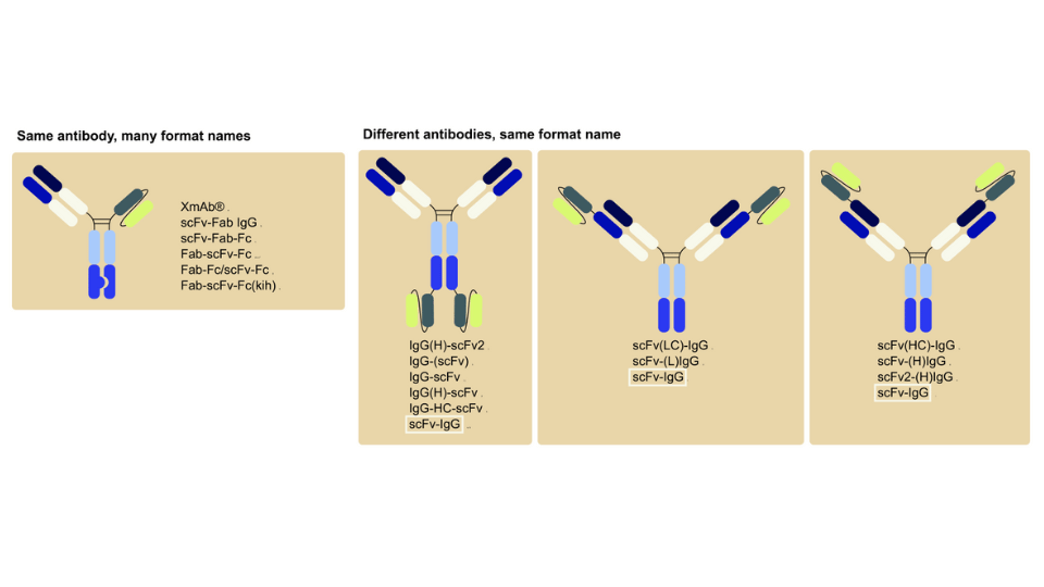
The versatility of cloning techniques and the variety of plasmids available allow the creation of wide varieties of antibody formats, ranging from full-length antibodies to antibody fragments and antibody-drug conjugates. The techniques and plasmids have become essential tools in antibody engineering and are likely to play a critical contribution to the development of future therapeutics.