Landscape of Camelid antibodies

Structural and functional adaptations, evolutionary origins and genomic organization in heavy chain antibodies.

Category:
Science
Date:
August 30, 2024
Read time:
10
min
Phylogenetic tree showcasing the evolutionary origins and distance between human, alpaca, rabbit and mouse , and visualization of main advantages of HCAbs as the small size, stable under temperature and chemical conditions, deep tissue penetration, recognition of distinct epitopes, and simple manufacturing.‍

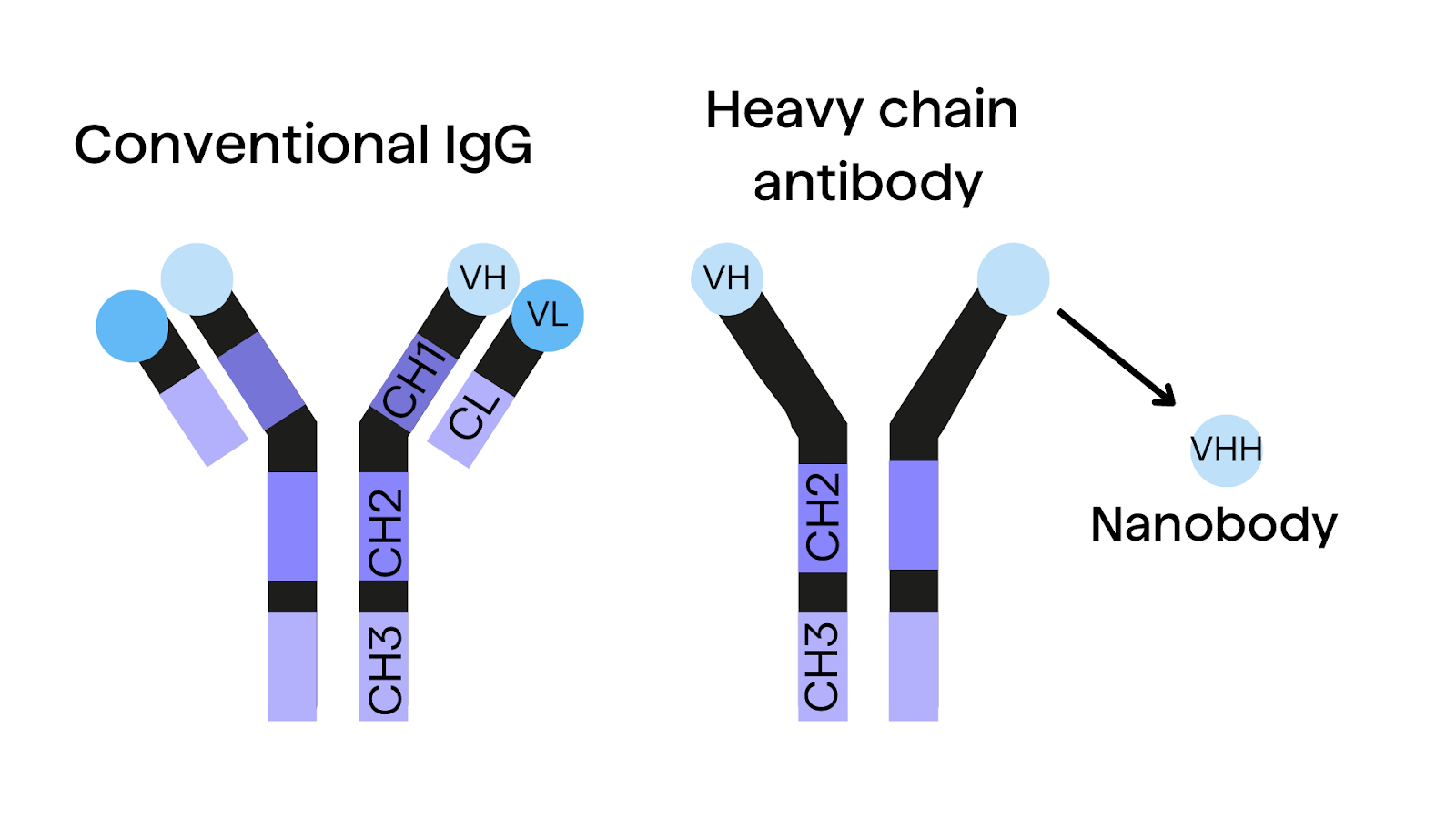
Camelids, including camels, llamas, and alpacas, possess a unique antibody system that has been gaining significant attention in recent years. These animals produce not only conventional antibodies but also a distinct class of antibodies known as heavy-chain antibodies (HCAbs), which lack light chains. The discovery of HCAbs led to the development of a new type of antibody fragments - single-domain antibodies or nanobodies. The properties of HCAbs and nanobodies have been very promising in the field of medicine and diagnostics, so the experiments around them continue.

Comparison of immunoglobulin structure of different antibodies: conventional IgGs, heavy chain antibodies (HCAbs) and nanobodies (VHHs) from camelids.
Figure 1. Comparison of conventional IgG antibody structure with heavy chain antibody and nanobody.

Evolutionary Origins and Advantages of Camelid antibodies

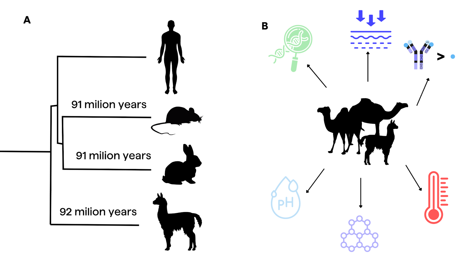
Camelids belong to the order Cetartiodactyla and share an evolutionary distance of approximately 92 million years from humans, comparable to the divergence between humans and mice5,6. This evolutionary distance has contributed to the development of unique antibody structures in camelids, such as HCAbs and nanobodies.

Nanobodies offer numerous advantages for laboratory and therapeutic applications. They are small (approximately 15 kDa ) in comparison to conventional IgGs (around 150 kDa)2,25, stable under a wide range of temperature and chemical conditions2, easy to clone2, and amenable to screening using phage display7. Additionally, camelid VHHs can recognize distinct epitopes that are inaccessible to human antibodies, as demonstrated in the case of SARS-CoV-2 nanobodies9. Their small size also simplifies humanization, manufacturing, and tissue penetration, although it can lead to rapid clearance and poor pharmacokinetic properties, which can be addressed by engineering them into conjugated or multivalent formats9.

Phylogenetic tree showcasing the evolutionary origins and distance between human, alpaca, rabbit and mouse , and visualization of main advantages of HCAbs as the small size, stable under temperature and chemical conditions, deep tissue penetration, recognition of distinct epitopes, and simple manufacturing.‍
Figure 2. A. Phylogenetic tree of divergent species, shown with respect to evolutionary distance from humans. B. Visualization of advantages of camelid antibodies: small size, stable under temperature and chemical conditions, deep tissue penetration, recognition of distinct epitopes, simple manufacturing.

Structural and Functional Adaptations of HCAbs

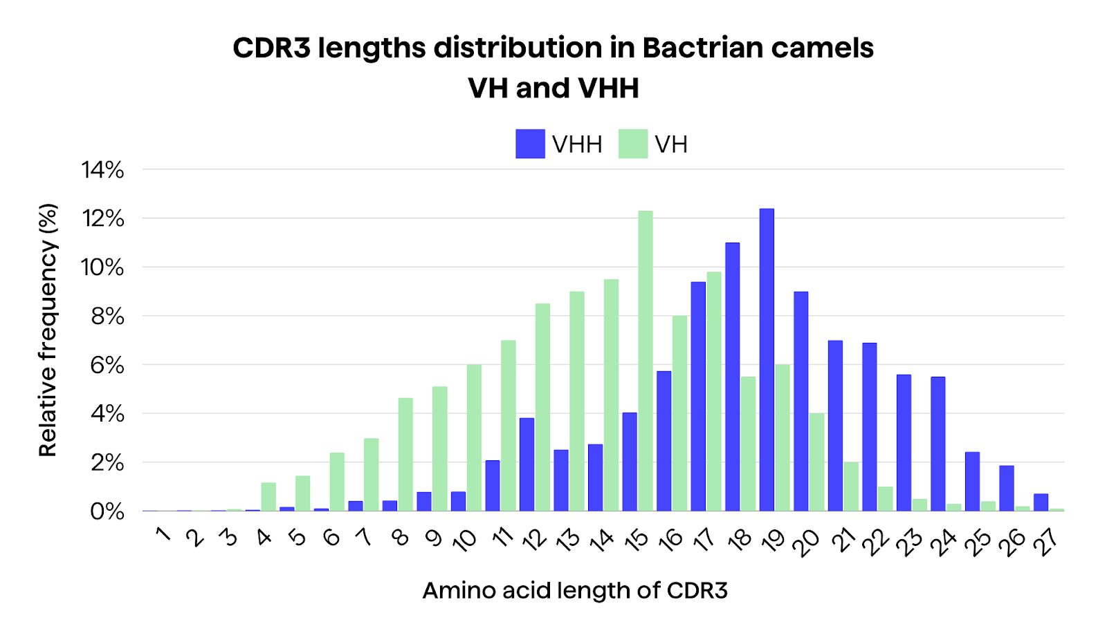
Some studies show differences between VH and VHH in camelid repertoires. In a comparative analysis from Li et al3 of the immune repertoire between VH and VHH in three Bactrian camels, HCAbs seems to exhibit longer CDR3 lengths, with an average of 18 amino acids compared to 13 amino acids in conventional antibodies3. This increase in CDR3 length again may enable access to sterically hindered sites, such as ligand-binding pockets and enzyme active sites3.

Bar chart presenting a comparison of CDR3 length distribution in Bactrian camels VH and VHH, pointing at the differences of relative frequency on each position in amino acid length of CDR3.
Figure 3. The CDR3 comparison of Bactrian camels including CDR3 length (AA) and proportion adapted from Li et al3.

Additionally, HCAbs demonstrated a higher mutation rate (12.3% vs. 8.3% in conventional antibodies) and characteristic amino acid substitutions, particularly at positions 49, 50, 52, 54, 57, 96, and 1013. These substitutions may likely contribute to the increased solubility and structural stability of HCAbs, compensating for the lack of a light chain3.

Box plots of the unique sequence numbers of each sub-region and diversity evaluation. a The unique number of sub-regions including FR1, CDR1, FR2, CDR2, FR3, CDR3, and FR4 in three samples. b Diversity evaluation using the normalized unique number by length, which equals to the unique number divided by average length of each sub-region. Box plot explanation: upper horizontal line of box, 75th percentile; lower horizontal line of box, 25th percentile; horizontal bar within box, the median of the three samples’ data; upper end of the whisker, maximum of the three samples’ data; lower end of the whisker, minimum of the three samples’ data.
Figure 4. Box plots of the unique sequence numbers of each sub-region and diversity evaluation from  Li et al3. A The unique number of sub-regions including FR1, CDR1, FR2, CDR2, FR3, CDR3, and FR4 in three samples. B Diversity evaluation using the normalized unique number by length, which equals the unique number divided by average length of each sub-region.

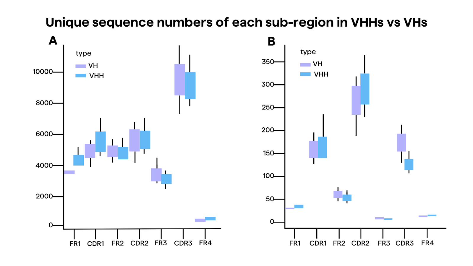
Furthermore, the distribution of cysteine codons, which form disulfide bonds to stabilize the antibody structure, seemed significantly higher in VHHs compared to VHs, especially in the CDR1 region (9.8% vs. 0.5%)3. This suggests that HCAbs may have evolved additional disulfide bonds to maintain structural integrity in the absence of a light chain.

Structural studies have revealed that the CDRs of camelid sdAbs can adopt a wide range of conformations, contributing to their ability to recognize diverse epitopes. Examples include intracellular targets10,11 or epitopes concealed from mAbs in protein structures12, G protein-coupled receptors13,14, and ion channels15.  For instance, the CDR3 loop can form extended or protruding structures that can access clefts or cavities on antigens, while the CDR1 loop can participate in antigen binding more extensively than in conventional antibodies. Additionally, the CDR2 loop often plays a crucial role in shaping the conformation of the CDR3 loop and determining antigen specificity.5

Non-Classical VHH Clones and Genomic Organization

While genomic and cDNA studies have shed light on the camelid VHH gene repertoire, a complete picture of the full germline V gene repertoires for both heavy and light chains is still lacking17. Preliminary investigations suggest that camelid VHH genes are highly homologous to the human VH3 family, with some key amino acid substitutions in the FR2.18 However, recent studies have also identified novel camelid V genes closely related to human IGHV5 and IGHV7 families.19 Ongoing efforts to fully characterize the camelid germline V gene repertoires will further our understanding of the evolutionary origins and diversification mechanisms of HCAbs.5

A subset of "non-classical" VHH clones, comprising approximately 8% of the total, are derived from conventional VH genes rather than the typical VHH genes16,17. These non-classical VHHs exhibit distinct features, such as a higher frequency of arginine substitution at the first position of the framework 4 region and shorter CDR3 lengths compared to classical VHHs3.

The genomic organization of the camelid IgH locus sheds light on the origins of conventional and homodimeric IgGs4. The IgH locus contains an intermixed arrangement of VHH genes, which encode the variable regions of homodimeric IgGs, and conventional VH genes, which encode the variable regions of tetrameric IgGs4. Downstream of the V gene cluster is a single DH-JH gene cluster, followed by the C gene region, which includes both CHH genes encoding the constant regions of homodimeric IgGs and conventional CH genes4.

A. Bar chart displaying the proportion of VHH and VH genes in eight Bactrian camels, with sample sizes ranging from 81 to 104 detected genes. The percentage of VHH genes ranges from approximately 21.69% to 37.93%, averaging at 29.78%. The VH genes consistently occupy a larger proportion compared to VHH genes across all samples. B. Phylogenetic tree depicting the relationships among IGHV gene subgroups (IGHV1, IGHV2, IGHV3) in camelids. The tree shows that the IGHV3 subgroup contains both VHH and VH genes, unlike the IGHV1 and IGHV2 subgroups which exclusively contain VH genes. The IGHV3 subgroup is further divided into distinct subsets with varying bootstrap values (62, 95, 100) indicating confidence levels in the branching patterns
Figure 5. Overview of IGHV3 gene repertoire in Camelids. A. The proportion of VHH and VH genes in eight Bactrian camels from Liu et al.26. The detected gene number ranged from 81 to 104 (with mean value of 90 ±9), and the proportion of VHH genes ranged from 21.69% to 37.93% (with mean value of 29.78% ±6.01%). B. Achour et al.27 found that the IgHV3, IgHV2, and IgHV1 subgroups contain 71, 11, and 6 V gene members, respectively. Interestingly, only the IgHV3 subgroup contains both VHH and VH genes (17 VHH members divided into six subsets and 54 VH members divided into 12 subsets; , whereas the IgHV1 and IgHV2 subgroups contain exclusively VH members. The numbers 62, 95, and 100 represent bootstrap values.  The length of branch on the bottom represents an amount genetic change of 0.087.

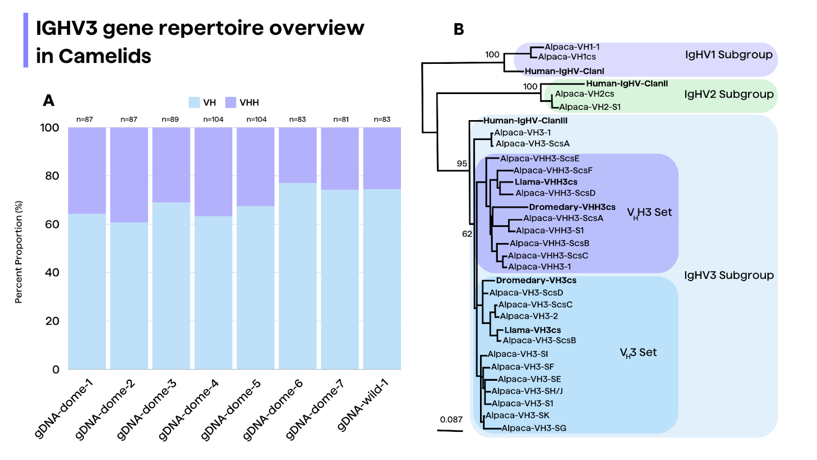
Phylogenetic analysis suggests that the VHH genes emerged from pre-existing VH genes of the IGHV3 subgroup, while the IGHV1 and IGHV2 subgroups contain only conventional VH genes.26 This supports a model in which homodimeric IgG-expressing B cells develop through an IgM+ stage, similar to the development of conventional IgG-expressing B cells26,27.

Therapeutic and Diagnostic Applications

The unique properties of camelid antibodies, particularly nanobodies, have opened up a wealth of opportunities for therapeutic and diagnostic applications. As of 2020, there were at least 37 camelid antibodies in clinical trials and numerous others in preclinical stages of development2.

Caplacizumab21, a bivalent nanobody was the first approved monoclonal antibody obtained from a non-rodent animal source2. It targets von Willebrand factor for the treatment of thrombotic thrombocytopenic purpura and thrombosis.22

Nanobodies have shown promise in targeting challenging and conserved drug targets, such as enzymes and G-protein-coupled receptors (GPCRs)20. Their long CDR3 regions enable them to access sterically hindered sites, making them attractive candidates for developing inhibitory, antagonist, and agonist antibodies against these targets2.

Furthermore, the ability of camelid antibodies to recognize distinct epitopes from human antibodies has been harnessed in the development of SARS-CoV-2 nanobodies that can bind to regions of the virus inaccessible to human MAbs23. This highlights the potential of camelid antibodies in addressing emerging infectious diseases and other challenging targets.

Future Directions and Concluding Remarks

The camelid antibody repertoire represents a valuable resource for antibody discovery and development, offering unique structural and functional properties that complement conventional antibody platforms. While camelids provide attractive features for antibody discovery, practical considerations such as the animal’s large size, limited accessible B cell repertoire, and mammalian genome homology to humans also present challenges2.

Additionally, the increasing availability of camelid genomic and transcriptomic data, coupled with advanced computational tools, will facilitate a deeper understanding of the evolutionary origins, diversification mechanisms, and structural adaptations of camelid antibodies3,4.

While camelid sdAbs offer several advantages over conventional mAbs and antibody fragments, their novel nature also presents unique regulatory challenges5. As the first therapeutic products of their kind, sdAb-based therapeutics will likely face increased scrutiny from regulatory agencies regarding their safety, immunogenicity, and manufacturing processes. Additionally, establishing standardized analytical and characterization methods for these single-domain antibodies may pose challenges, as they differ structurally from conventional mAbs. Overcoming these regulatory hurdles will be crucial for the successful development and approval of sdAb-based therapeutics5.

References

Right-pointing black chevron
  1. Muyldermans S, Baral TN, Retamozzo VC, De Baetselier P, De Genst E, Kinne J, Leonhardt H, Magez S, Nguyen VK, Revets H, Rothbauer U, Stijlemans B, Tillib S, Wernery U, Wyns L, Hassanzadeh-Ghassabeh G, Saerens D. Camelid immunoglobulins and nanobody technology. Vet Immunol Immunopathol. 2009 Mar 15;128(1-3):178-83. doi: 10.1016/j.vetimm.2008.10.299. Epub 2008 Oct 17. PMID: 19026455.
  2. Banik SSR, Kushnir N, Doranz BJ, Chambers R. Breaking barriers in antibody discovery: harnessing divergent species for accessing difficult and conserved drug targets. MAbs. 2023 Jan-Dec;15(1):2273018. doi: 10.1080/19420862.2023.2273018. Epub 2023 Dec 5. PMID: 38050985; PMCID: PMC10793698.
  3. Li X, Duan X, Yang K, Zhang W, Zhang C, Fu L, Ren Z, Wang C, Wu J, Lu R, Ye Y, He M, Nie C, Yang N, Wang J, Yang H, Liu X, Tan W. Comparative Analysis of Immune Repertoires between Bactrian Camel's Conventional and Heavy-Chain Antibodies. PLoS One. 2016 Sep 2;11(9):e0161801. doi: 10.1371/journal.pone.0161801. PMID: 27588755; PMCID: PMC5010241.
  4. Bactrian Camels Genome Sequencing and Analysis Consortium; Jirimutu, Wang Z, Ding G, Chen G, Sun Y, Sun Z, Zhang H, Wang L, Hasi S, Zhang Y, Li J, Shi Y, Xu Z, He C, Yu S, Li S, Zhang W, Batmunkh M, Ts B, Narenbatu, Unierhu, Bat-Ireedui S, Gao H, Baysgalan B, Li Q, Jia Z, Turigenbayila, Subudenggerile, Narenmanduhu, Wang Z, Wang J, Pan L, Chen Y, Ganerdene Y, Dabxilt, Erdemt, Altansha, Altansukh, Liu T, Cao M, Aruuntsever, Bayart, Hosblig, He F, Zha-ti A, Zheng G, Qiu F, Sun Z, Zhao L, Zhao W, Liu B, Li C, Chen Y, Tang X, Guo C, Liu W, Ming L, Temuulen, Cui A, Li Y, Gao J, Li J, Wurentaodi, Niu S, Sun T, Zhai Z, Zhang M, Chen C, Baldan T, Bayaer T, Li Y, Meng H. Genome sequences of wild and domestic bactrian camels. Nat Commun. 2012;3:1202. doi: 10.1038/ncomms2192. Erratum in: Nat Commun. 2013;4. doi: 10.1038/ncomms3089. PMID: 23149746; PMCID: PMC3514880.
  5. Achour I, Cavelier P, Tichit M, Bouchier C, Lafaye P, Rougeon F. Tetrameric and homodimeric camelid IgGs originate from the same IgH locus. J Immunol. 2008 Aug 1;181(3):2001-9. doi: 10.4049/jimmunol.181.3.2001. PMID: 18641337.
  6. Hedges SB. The origin and evolution of model organisms. Nat Rev Genet. 2002;3:838–49. doi:10.1038/nrg929. PMID: 12415314.
  7. International Chicken Genome Sequencing C. Sequence and comparative analysis of the chicken genome provide unique perspectives on vertebrate evolution. Nature. 2004;432:695–716. PMID: 15592404. doi:10.1038/nature03154
  8. Marks JD, Bradbury A. Selection of human antibodies from phage display libraries. Methods Mol Biol. 2004;248:161–76. doi:10.1385/1-59259-666-5:161. PMID: 14970495.
  9. Xiu J, Xu K, Jung S, Conte A, Lieberman J, Muecksch F, Lorenzi JCC, Park S, Schmidt F, Wang Z, et al. Nanobodies from camelid mice and llamas neutralize SARS-CoV-2 variants. Nature. 2021;595:278–82. doi:10.1038/s41586-021-03676-z. PMID: 34098567.
  10. Jovcevska I, Muyldermans S. 2020. The therapeutic potential of nanobodies. BioDrugs: clinical immunotherapeutics, biopharmaceuticals and gene therapy. PMID: 31686399 34(1):11–26. doi:10.1007/s40259-019-00392-z
  11. McGonigal K, Tanha J, Palazov E, Li S, Gueorguieva-Owens D, Pandey S. Isolation and functional characterization of single domain antibody modulators of caspase-3 and apoptosis. Appl Biochem Biotechnol (2009) 157:226–36. 10.1007/s12010-008-8266-4
  12. Staus DP, Wingler LM, Strachan RT, Rasmussen SG, Pardon E, Ahn S, et al. Regulation of β2-adrenergic receptor function by conformationally selective single-domain intrabodies. Mol Pharmacol (2014) 85:472–81. 10.1124/mol.113.089516
  13. Stijlemans B, Conrath K, Cortez-Retamozo V, Van Xong H, Wyns L, Senter P, et al. Efficient targeting of conserved cryptic epitopes of infectious agents by single domain antibodies: African trypanosomes as paradigm. J Biol Chem (2004) 279:1256–61. 10.1074/jbc.M307341200
  14. Bradley ME, Dombrecht B, Manini J, Willis J, Vlerick D, De Taeye S, et al. Potent and efficacious inhibition of CXCR2 signaling by biparatopic nanobodies combining two distinct modes of action. Mol Pharmacol (2015) 87:251–62. 10.1124/mol.114.094821
  15. Manglik A, Kobilka BK, Steyaert J. Nanobodies to study G protein-coupled receptor structure and function. Annu Rev Pharmacol Toxicol (2017) 57:19–37. 10.1146/annurev-pharmtox-010716-104710
  16. Muyldermans S. Nanobodies: natural single-domain antibodies. Annu Rev Biochem (2013) 82:775–97. 10.1146/annurev-biochem-063011-092449
  17. Griffin LM, Snowden JR, Lawson AD, Wernery U, Kinne J, Baker TS. Analysis of heavy and light chain sequences of conventional camelid antibodies from Camelus dromedarius and Camelus bactrianus species. Journal of immunological methods. 2014; 405:35–46. Epub 2014/01/22. doi: 10.1016/j.jim.2014.01.003 PMID: 24444705.
  18. Vu KB, Ghahroudi MA, Wyns L, Muyldermans S. Comparison of llama VH sequences from conventional and heavy chain antibodies. Mol Immunol (1997) 34:1121–31. 10.1016/S0161-5890(97)00146-6
  19. Klarenbeek A, El Mazouari K, Desmyter A, Blanchetot C, Hultberg A, de Jonge N, et al. Camelid Ig V genes reveal significant human homology not seen in therapeutic target genes, providing for a powerful therapeutic antibody platform. MAbs (2015) 7:693–706. 10.1080/19420862.2015.1046648
  20. Klarenbeek A, El Mazouari K, Desmyter A, Blanchetot C, Hultberg A, de Jonge N, et al. Camelid Ig V genes reveal significant human homology not seen in therapeutic target genes, providing for a powerful therapeutic antibody platform. MAbs (2015) 7:693–706. 10.1080/19420862.2015.1046648
  21. Heukers R, De Groof TWM, Smit MJ. Nanobodies detecting and modulating GPCRs outside in and inside out. Curr Opin Cell Biol. 2019;57:115–22. doi:10.1016/j.ceb.2019.01.003. PMID: 30849632
  22. Alfaleh MA, Alsaab HO, Mahmoud AB, Alkayyal AA, Jones ML, Mahler SM, Hashem AM. Phage display derived monoclonal antibodies: from bench to bedside. Front Immunol. 2020;11:1986. doi:10.3389/fimmu.2020.01986. PMID: 32983137.
  23. Bowyer A, Brown P, Hopkins B, Scully M, Shepherd F, Lowe A, Mensah P, Maclean R, Kitchen S, van Veen JJ. Von Willebrand factor assays in patients with acquired immune thrombotic thrombocytopenia purpura treated with caplacizumab. Br J Haematol. 2022 May;197(3):349-358. doi: 10.1111/bjh.18080. Epub 2022 Mar 8. PMID: 35262910.
  24. Barnes CO, Jette CA, Abernathy ME, Dam KA, Esswein SR, Gristick HB, Malyutin AG, Sharaf NG, Huey-Tubman KE, Lee YE, et al. SARS-CoV-2 neutralizing antibody structures inform therapeutic strategies. Nature. 2020;588(7839):682–87. PMID:33045718. doi:10.1038/s41586-020-2852-1.
  25. Zhang Q, Zhang N, Xiao H, Wang C, He L. Small Antibodies with Big Applications: Nanobody-Based Cancer Diagnostics and Therapeutics. Cancers (Basel). 2023 Nov 29;15(23):5639. doi: 10.3390/cancers15235639. PMID: 38067344; PMCID: PMC10705070.
  26. Liu Y, Yi L, Li Y, Wang Z, Jirimutu. Characterization of heavy-chain antibody gene repertoires in Bactrian camels. J Genet Genomics. 2023 Jan;50(1):38-45. doi: 10.1016/j.jgg.2022.04.010. Epub 2022 Apr 29. PMID: 35500746.
  27. Ikbel Achour, Patricia Cavelier, Magali Tichit, Christiane Bouchier, Pierre Lafaye, François Rougeon; Tetrameric and Homodimeric Camelid IgGs Originate from the Same IgH Locus. J Immunol 1 August 2008; 181 (3): 2001–2009. https://doi.org/10.4049/jimmunol.181.3.2001

Annotate the VHH with two clicks!

Other recent posts